

What is Flow Imaging Microscopy (FIM)?
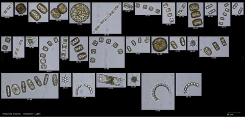
Flow Imaging Microscopy is a rapid, automated technique that captures high-resolution digital images of microscopic particles suspended in a liquid. With the FlowCam system, users can quickly assess the size, shape, number and identity of particles within a sample.
The technique was first developed at the Bigelow Laboratory for Ocean Sciences in Boothbay Harbour, Maine. At the time, researchers relied on labour-intensive methods like traditional microscopy and flow cytometry to identify and count plankton. To improve this, Bigelow scientists designed the original FlowCam, combining the advantages of both tools into a single instrument. With FlowCam, ocean water samples could be digitally imaged and analysed in real time.
Modern Applications Across Industries
Today, FlowCam is widely used in laboratories that require detailed particle characterisation across a variety of sectors, including:
-
Aquatic sciences: Studying microorganisms in marine and freshwater environments to understand key ecological processes.
-
Biopharmaceuticals: Monitoring subvisible particles and protein aggregates in injectable drugs to assess formulation stability.
-
Cell and gene therapies: Analysing cells, drug carriers, and delivery particles in advanced therapeutic products.
-
Food and beverage: Ensuring ingredient quality where particle shape impacts texture and taste.
-
Materials science: Supporting product development and quality control for polymers, fibres, microspheres, and emulsions.
How Flow Imaging Particle Analysis Works
The FlowCam streamlines particle analysis. A liquid sample is introduced into a flow cell and passes between a light source and a microscope objective aligned with a digital camera. As particles flow through this path, the camera captures images—up to 50,000 particles per minute.
FlowCam’s proprietary VisualSpreadsheet software processes these images in real time, isolating and measuring individual particles. It records key parameters such as count, diameter, volume, and aspect ratio, along with advanced shape descriptors like circularity, elongation, perimeter, and even features like transparency and colour. The data can be filtered, sorted, classified and displayed in multiple formats for detailed interpretation.
Why Flow Imaging Offers Greater Accuracy
A major advantage of using FlowCam is that particle measurements are taken directly from each particle’s image. With fixed optics and known magnifications, distances on the image can be accurately converted to real-world sizes—no assumptions required.
By contrast, other analytical techniques such as light obscuration, laser diffraction or dynamic light scattering rely on indirect measurements. They infer size and shape based on how light interacts with a population of particles, often assuming a particular geometry or averaging out individual characteristics.
Seeing Particles, Not Just Numbers
Unlike traditional methods that assess bulk properties, flow imaging microscopy allows you to visualise and analyse individual particles. This gives a clearer, more accurate picture of your sample and provides valuable insight into the behaviour and characteristics of particles—critical for research, development and quality control.
With FlowCam, you gain the ability to directly observe your sample, take precise measurements, and extract meaningful data—all in one streamlined, efficient process.
For more information on What is Flow Imaging Microscopy (FIM)? talk to Meritics Ltd